

Introduction to Oxidative Stress During Embryonic Development
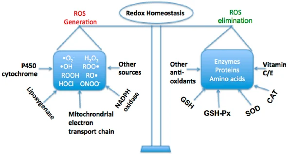
Oxidative stress plays a central role in embryonic growth, cellular signaling, tissue formation, and organ development. During embryogenesis, the balance between oxidants and antioxidants regulates many biological mechanisms that determine whether embryonic cells proliferate, differentiate, survive, or undergo programmed cell death. Reactive oxygen species (ROS), once considered only harmful metabolic byproducts, are now recognized as essential signaling molecules involved in normal developmental pathways.
Embryonic development requires tightly controlled redox regulation. Redox status influences mitochondrial activity, gene transcription, protein signaling, stem cell differentiation, and apoptosis. Controlled oxidative signaling supports normal morphogenesis, while excessive oxidative stress can disrupt embryonic development and lead to congenital abnormalities, developmental delay, embryotoxicity, or fetal death.
Research in developmental biology demonstrates that ROS are required for several physiological embryonic processes, including blastocyst formation, neural differentiation, vascular development, limb patterning, and tissue remodeling. However, uncontrolled accumulation of ROS damages lipids, proteins, DNA, and cellular membranes, creating oxidative injury that interferes with normal fetal growth.
This complex relationship shows that embryonic development depends on a carefully regulated oxidative environment in which both oxidative stress and antioxidant defense systems must remain balanced.
Reactive Oxygen Species and Cellular Redox Regulation
Formation of Reactive Oxygen Species
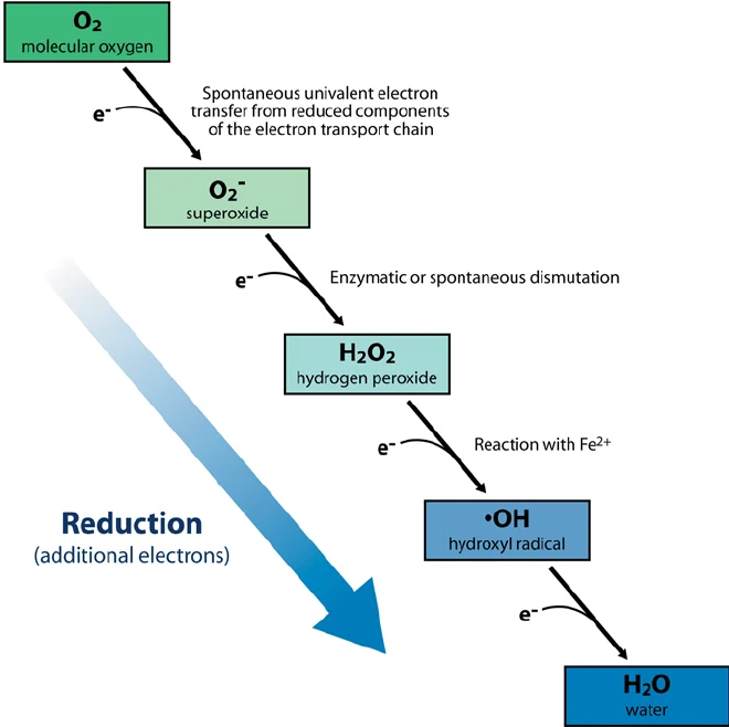
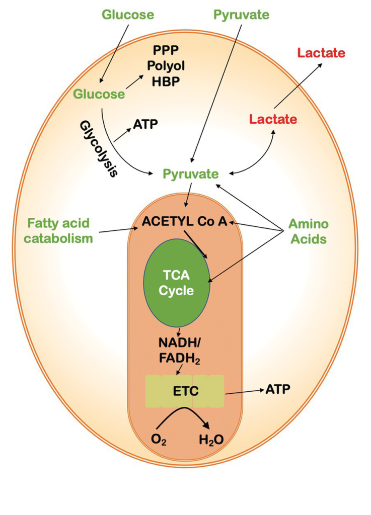
Reactive oxygen species are generated mainly through mitochondrial oxidative metabolism. During ATP production in the electron transport chain, oxygen molecules may undergo incomplete reduction, leading to the formation of several ROS types, including:
- Superoxide anion
- Hydrogen peroxide
- Hydroxyl radicals
Although mitochondrial respiration is highly efficient, small amounts of electrons leak from respiratory complexes, creating oxidative molecules capable of influencing cellular activity.
ROS can also be generated through:
- Growth factor signaling
- Inflammatory pathways
- Xenobiotic metabolism
- Environmental toxins
- Cellular stress responses
Unlike traditional assumptions that ROS are exclusively toxic, modern embryology research confirms that ROS act as intracellular signaling mediators. Their rapid formation and degradation allow them to regulate multiple developmental pathways with high precision.
Physiological Role of Oxidative Stress in Normal Embryonic Development
Redox Balance Controls Cellular Fate
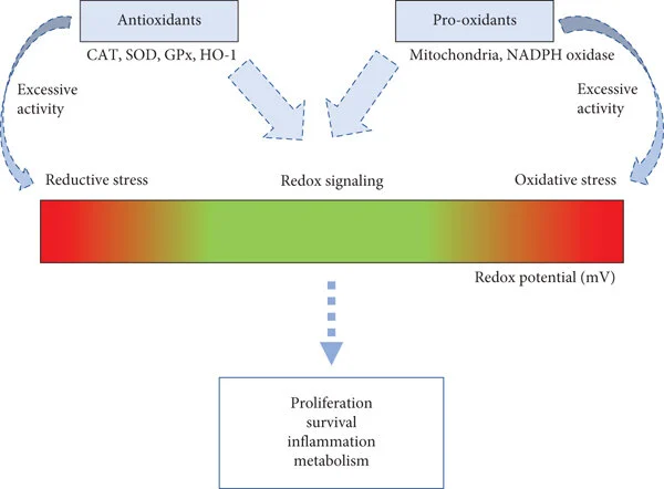
The oxidative environment within embryonic tissues directly determines cellular behavior. Different redox states guide embryonic cells toward distinct biological outcomes:
- Highly reduced conditions favor cell proliferation
- Mild oxidative conditions promote differentiation
- Strong oxidative stress induces apoptosis or necrosis
This process is commonly referred to as redox switching, a mechanism essential for developmental timing and tissue organization.
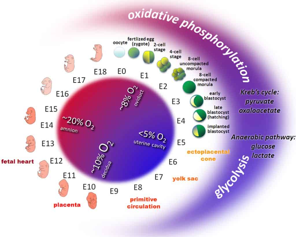
During early embryogenesis, the intrauterine environment remains relatively hypoxic. Low oxygen levels protect the embryo during organogenesis, when tissues are highly sensitive to oxidative damage. Later in development, establishment of placental circulation increases oxygen exposure, requiring stronger antioxidant defenses to maintain redox homeostasis.
Oxidative Stress and Embryonic Cell Differentiation
ROS-Mediated Differentiation Mechanisms
Several studies demonstrate that controlled oxidative stress promotes embryonic cell differentiation.
Examples include:
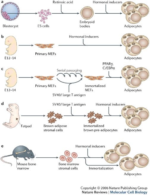
Adipocyte Differentiation
Undifferentiated preadipocytes contain elevated levels of reduced glutathione (GSH). During differentiation, oxidized glutathione (GSSG) levels rise significantly, indicating that oxidative changes trigger maturation processes.
Bone and Odontoblast Development
Low concentrations of hydrogen peroxide stimulate:
- Alkaline phosphatase activity
- Mineralized matrix formation
- Osteoblast differentiation
These markers indicate that moderate ROS exposure supports skeletal tissue formation.
Neuronal Development
In chick embryo models, antioxidants prevented programmed neuronal death. However, excessive antioxidant exposure was equally harmful, confirming that embryonic tissues require a precise oxidative balance for proper neurodevelopment.
Oxidative Stress and Embryonic Gene Regulation
Redox-Sensitive Transcription Factors
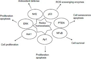
Reactive oxygen species regulate numerous transcription factors involved in embryonic development. These transcriptional regulators influence angiogenesis, organogenesis, cell survival, and apoptosis.
Major redox-sensitive transcription factors include:
- HIF-1
- NF-kB
- AP-1
- Ref-1
- Nrf1
- Wnt/beta-catenin signaling
These molecular systems connect oxidative status with embryonic gene expression.
HIF-1 and Hypoxia Signaling During Embryogenesis
Role of HIF-1 in Vascular and Neural Development
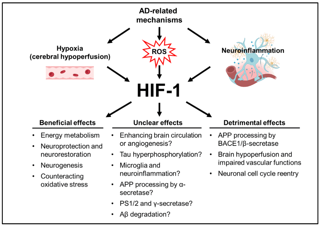
Hypoxia-inducible factor 1 (HIF-1) regulates genes responsible for:
- Angiogenesis
- Energy metabolism
- Erythropoiesis
- Cell survival
HIF-1 activation occurs under low oxygen conditions and stimulates vascular endothelial growth factor (VEGF) expression, promoting blood vessel formation.
Loss of HIF-1 function causes:
- Cardiac malformations
- Neural defects
- Abnormal vascular development
- Embryonic lethality
HIF-1 is also essential for fetal iron regulation and embryonic erythropoiesis, emphasizing the importance of redox signaling during circulatory development.
NF-kB Signaling and Oxidative Stress
Developmental Functions of NF-kB
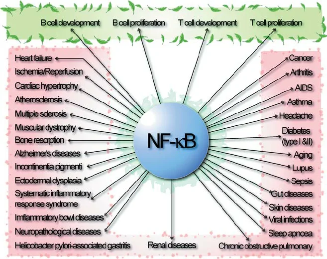
NF-kB regulates genes involved in:
- Inflammation
- Cell proliferation
- Anti-apoptotic signaling
- Cytokine expression
During fetal lung development, NF-kB signaling supports:
- Lung mesenchymal proliferation
- Sodium transport
- Fluid clearance
- Resistance to oxidative injury
Oxidative stress can activate NF-kB signaling pathways, potentially modifying embryonic development during maternal inflammation or environmental stress exposure.
AP-1 Transcription Factor and Developmental Oxidative Stress
AP-1 Activation During Organogenesis
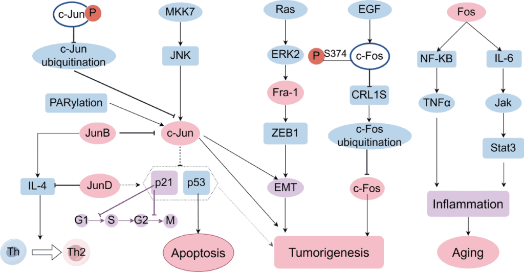
Activator protein-1 (AP-1) is one of the most important oxidative stress-responsive transcription factors.
During mid-organogenesis, AP-1 proteins increase significantly while the embryo undergoes rapid growth and differentiation. Excessive AP-1 activation caused by oxidative imbalance may lead to:
- Limb abnormalities
- Craniofacial defects
- Developmental arrest
- Increased apoptosis
Experimental depletion of glutathione during rat embryogenesis produced severe developmental abnormalities through increased AP-1 activity.
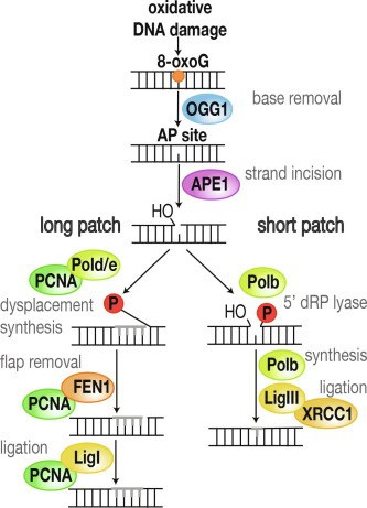
Ref-1 and DNA Repair During Embryonic Growth
Oxidative DNA Protection Mechanisms
Ref-1 is a multifunctional protein involved in:
- DNA repair
- Transcription factor regulation
- Oxidative stress response
Ref-1 maintains AP-1 activity by reducing oxidized transcription factors and repairing oxidative DNA lesions.
Loss of Ref-1 function results in:
- Extreme oxidative sensitivity
- Failed embryonic development
- Early embryonic death
These findings highlight the importance of oxidative DNA repair systems during rapid embryonic cell division.
Nrf1 and Antioxidant Gene Expression
Redox Homeostasis in Fetal Liver Development
Nrf1 regulates antioxidant defense genes that protect embryonic tissues from oxidative injury.
Nrf1 deficiency causes:
- Severe fetal anemia
- Impaired erythropoiesis
- Increased apoptosis
- Oxidative liver damage
This transcription factor is particularly important in maintaining redox balance during fetal liver maturation.
Wnt/Beta-Catenin Signaling and Oxidative Regulation
Oxidative Modulation of Embryonic Patterning
The Wnt/beta-catenin pathway controls:
- Organogenesis
- Tissue patterning
- Stem cell differentiation
- Morphogenesis
Oxidative stress negatively regulates Wnt signaling by reducing nuclear beta-catenin accumulation.
Hydrogen peroxide exposure suppresses Wnt target genes, potentially disrupting developmental processes such as neural tube formation and limb development.
Oxidative Stress and Abnormal Embryonic Development
Embryotoxicity and Teratogenesis
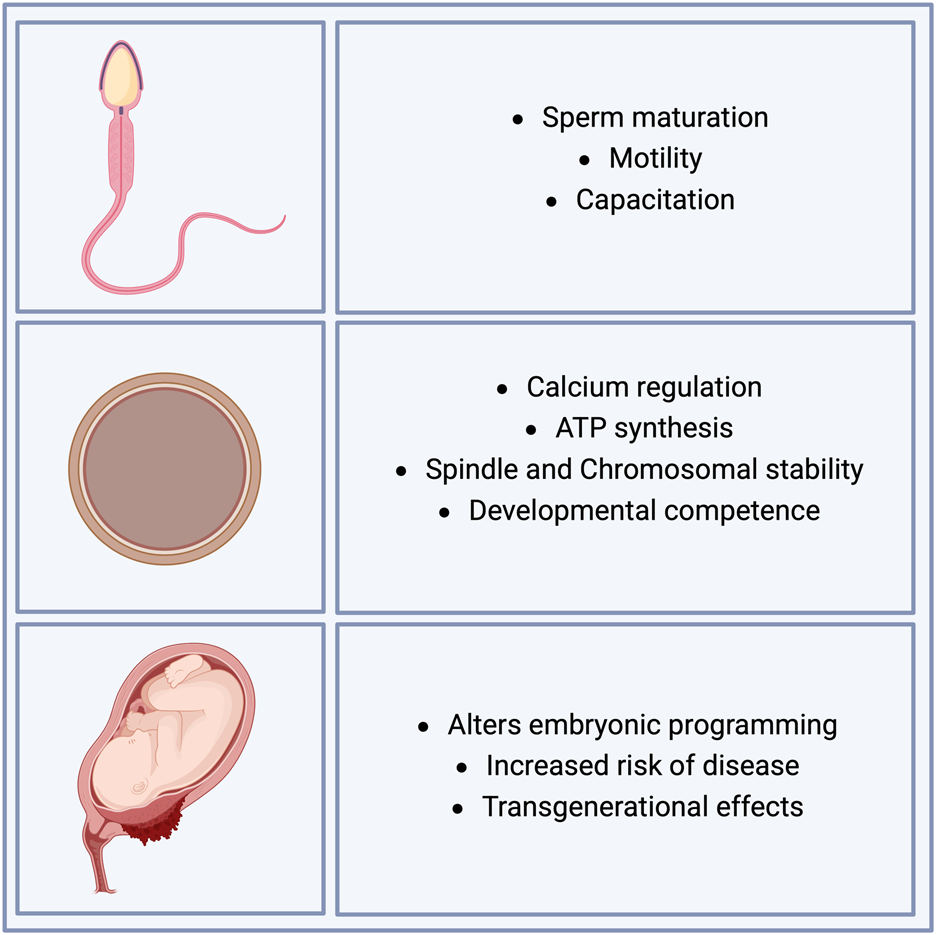
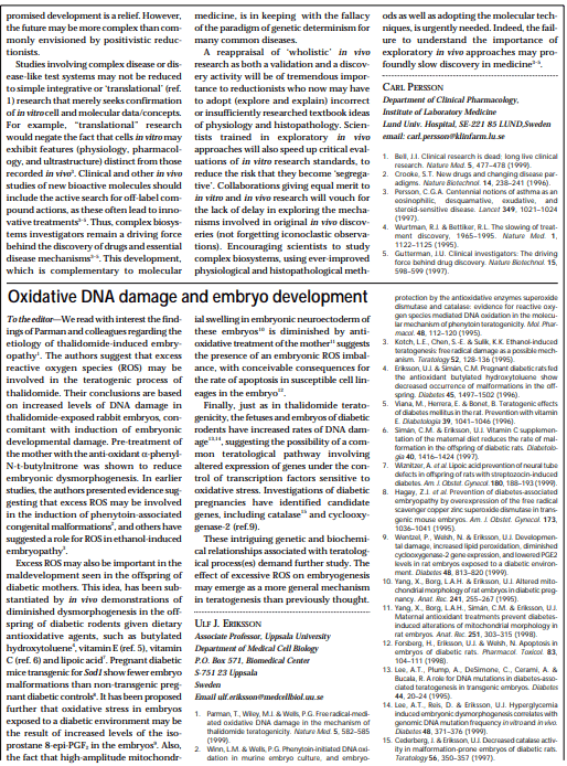
Although physiological ROS levels are required for development, excessive oxidative stress is strongly associated with embryotoxicity and congenital malformations.
Embryonic vulnerability depends on developmental stage, oxygen availability, and antioxidant capacity.
Major oxidative stress-related teratogens include:
- Thalidomide
- Phenytoin
- Ethanol
- Hyperglycemia associated with maternal diabetes
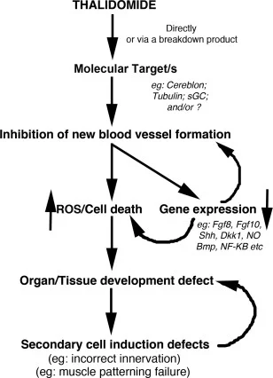
Thalidomide and Oxidative Embryopathy
Redox Imbalance and Limb Malformations
Thalidomide exposure is linked to severe congenital defects including:
- Phocomelia
- Limb reduction abnormalities
- Organ malformations
Research demonstrates that thalidomide induces oxidative stress through:
- Glutathione depletion
- Increased oxidized glutathione
- Nuclear redox imbalance
These alterations affect transcription factor signaling and embryonic developmental pathways.
Phenytoin-Induced Oxidative Stress
Oxidative DNA Damage During Embryogenesis
Phenytoin exposure during pregnancy increases the incidence of congenital abnormalities.
Mechanisms include:
- Oxidative DNA injury
- Hypoxia
- Vascular disruption
- NF-kB activation
Antioxidant supplementation significantly reduces phenytoin-associated embryopathies, confirming the role of ROS in developmental toxicity.
Ethanol and Fetal Oxidative Damage
Oxidative Mechanisms in Fetal Alcohol Toxicity
Alcohol metabolism generates large amounts of ROS and decreases antioxidant defenses.
Ethanol exposure during pregnancy causes:
- Lipid peroxidation
- Reduced glutathione levels
- Altered NF-kB signaling
- Increased embryonic oxidative stress
Antioxidants such as vitamin C and lipoic acid partially reduce ethanol-induced embryotoxicity.
Maternal Diabetes and Oxidative Embryopathy
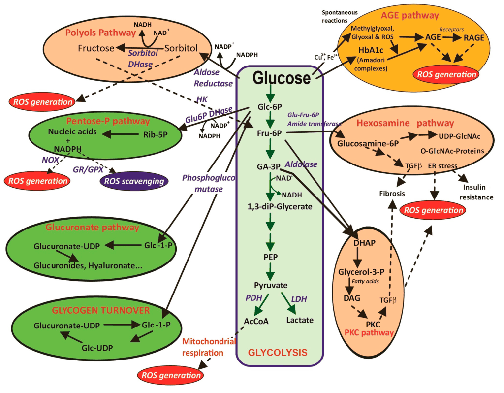
Hyperglycemia-Induced Oxidative Stress
Maternal diabetes is one of the most important causes of oxidative stress during pregnancy.
High glucose levels increase mitochondrial ROS production, resulting in:
- Neural tube defects
- Developmental abnormalities
- Reduced fetal growth
- Altered gene expression
Oxidative stress suppresses the developmental transcription factor Pax3, which is essential for neural crest migration and neural tube closure.
Antioxidant therapy using glutathione precursors and superoxide scavengers significantly reduces diabetic embryopathy.
Antioxidant Defense Systems During Embryonic Development
Glutathione and Embryonic Protection
Glutathione represents the major intracellular antioxidant system during embryogenesis.
Functions include:
- ROS detoxification
- Maintenance of redox balance
- Protection against DNA damage
- Regulation of signaling pathways
Deficiency in glutathione metabolism increases embryonic sensitivity to oxidative stress and congenital abnormalities.
Selenium and Selenoproteins in Embryogenesis
Developmental Role of Selenium-Dependent Antioxidants
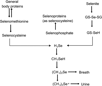
Selenoproteins are critical for antioxidant defense and embryonic survival.
Important selenoproteins include:
- Thioredoxin
- GPX4
- Selenoprotein W
Loss of these proteins results in:
- Early embryonic lethality
- Reduced cell proliferation
- Increased oxidative injury
- Defective tissue organization
Selenium-dependent enzymes are particularly important during neural, cardiac, and muscular development.
Lipoic Acid and Mitochondrial Antioxidant Defense
Role in Early Embryonic Metabolism
Lipoic acid functions as:
- A mitochondrial cofactor
- A potent antioxidant
- A regulator of cellular energy metabolism
Deficiency in lipoic acid synthesis causes severe embryonic developmental delay and embryonic death before mid-gestation.
G6PD and Embryonic Oxidative Resistance
Pentose Phosphate Pathway and Redox Protection
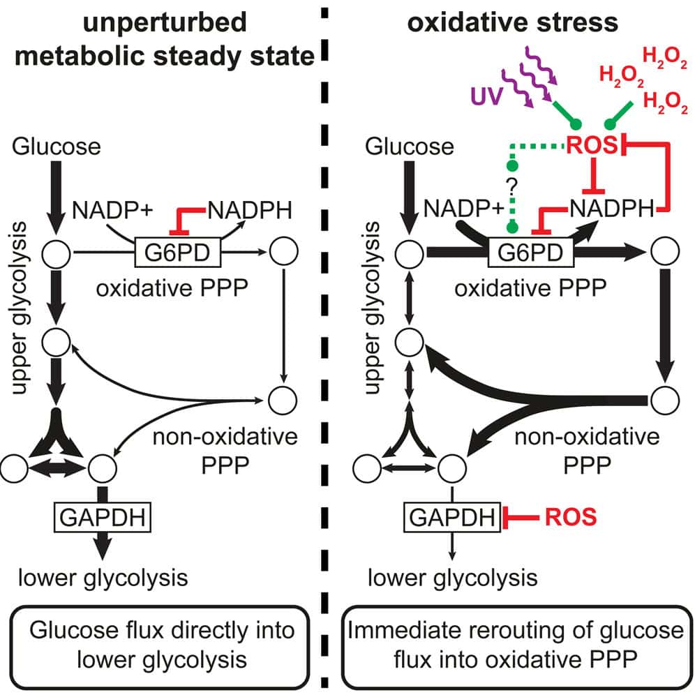
Glucose-6-phosphate dehydrogenase (G6PD) is essential for maintaining NADPH production and glutathione recycling.
G6PD deficiency increases:
- Embryonic oxidative stress
- DNA damage
- Fetal mortality
- Congenital defects
This enzyme plays a major protective role against endogenous and xenobiotic-induced oxidative injury.
Conclusion
Oxidative stress is a major regulatory mechanism in embryonic development. Reactive oxygen species influence proliferation, differentiation, apoptosis, gene expression, and organogenesis through highly controlled redox-dependent signaling pathways.
Physiological ROS levels are essential for normal embryogenesis, while excessive oxidative stress disrupts developmental processes and increases the risk of congenital malformations. Transcription factors such as HIF-1, NF-kB, AP-1, Nrf1, and Wnt signaling components are strongly influenced by cellular redox balance and contribute to embryonic patterning and tissue specification.
Environmental toxins, maternal diabetes, pharmaceutical agents, and metabolic disorders can disturb embryonic oxidative homeostasis and lead to developmental abnormalities. Antioxidant defense systems including glutathione, selenium-dependent enzymes, thioredoxin, and lipoic acid are critical for protecting embryonic tissues against oxidative damage.
Understanding oxidative stress biology during embryogenesis is essential for improving reproductive medicine, developmental toxicology, fetal health, stem cell biology, and prenatal therapeutic strategies.