
Introduction to Genitourinary Development
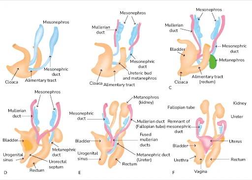
In mammals such as humans and mice, reproduction depends on internal fertilization, a biological process supported by the formation of specialized reproductive organs collectively known as the external genitalia. These structures originate during embryonic development from a transient embryological structure called the genital tubercle (GT), which represents the precursor of the penis in males and the clitoris in females.
The genital tubercle develops as a ventro-caudal embryonic structure composed primarily of intermediate mesoderm-derived mesenchymal cells surrounding a central endoderm-derived epithelial tube. During early embryogenesis, the genitalia of males and females are morphologically indistinguishable. Sexual differentiation begins later in development, around day 50 of human gestation and embryonic day 14–15 in mice, when sex-specific anatomical characteristics gradually emerge.
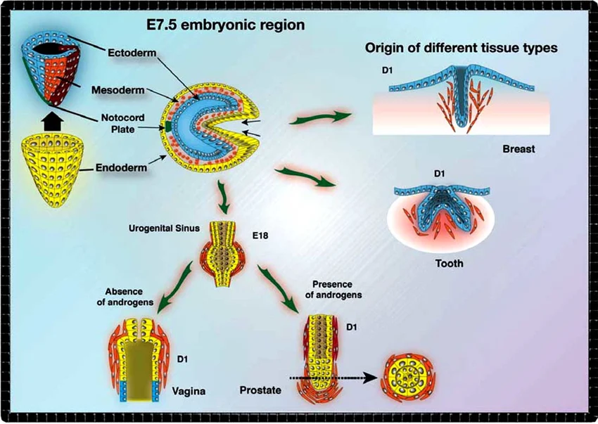
Historically, scientists believed that differentiation of the external genitalia was controlled mainly by androgen signaling. The presence of androgens drives male genital development, while the absence of androgen activity promotes female genital formation. However, modern developmental biology studies demonstrate that genitourinary development is regulated by a much more complex genetic and molecular network involving transcription factors, signaling pathways, morphogens, and epigenetic regulators.
Among the most important regulators of external genital formation are the homeobox transcription factors HOXA13 and HOXD13. These genes play essential roles in the regulation of tissue growth, epithelial-mesenchymal interactions, cell proliferation, apoptosis, and structural patterning during genitourinary morphogenesis.
Molecular Regulation of Genital Tubercle Development
Growth Factors Involved in Genitourinary Organogenesis
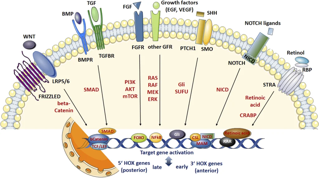
Development of the genital tubercle requires precise coordination between cellular proliferation, differentiation, migration, and programmed cell death. Multiple signaling molecules are involved in these processes, including:
- Fibroblast Growth Factor 8 (FGF8)
- Fibroblast Growth Factor 10 (FGF10)
- Sonic Hedgehog (SHH)
- Bone Morphogenetic Proteins BMP2, BMP4, and BMP7
These signaling pathways regulate:
- Tissue polarity
- Epithelial signaling
- Mesenchymal growth
- Morphogenetic remodeling
- Urethral closure
- External genital patterning
A major source of these signaling molecules is the centralized genital tubercle epithelium, which functions as an important developmental signaling center. This epithelial region supplies proliferative and positional signals that guide the surrounding mesenchymal tissue during genital formation.
Research has shown that disruption of these signaling mechanisms can result in severe congenital abnormalities affecting the urinary and reproductive systems.
HOX Genes and Developmental Regulation
Overview of HOX Gene Function
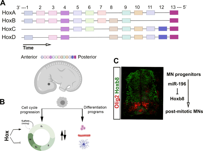
HOX genes encode a highly conserved family of transcription factors that regulate embryonic body patterning and organ development. In mammals, HOX genes are organized into four chromosomal clusters:
- HOXA
- HOXB
- HOXC
- HOXD
Together, these clusters contain 39 genes distributed across specific chromosomes in humans and mice.
HOX proteins function by binding DNA regulatory regions and controlling the expression of downstream developmental genes involved in:
- Organogenesis
- Skeletal patterning
- Limb development
- Urogenital differentiation
- Tissue polarity
- Cell fate specification
Among all HOX genes, HOXA13 and HOXD13 are particularly important for distal limb and genitourinary development.
Role of HOXA13 in Genitourinary Development
Hand-Foot-Genital Syndrome (HFGS)
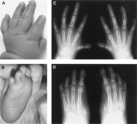
Mutations affecting HOXA13 are responsible for a rare autosomal dominant disorder known as Hand-Foot-Genital Syndrome (HFGS).
This syndrome causes severe developmental abnormalities involving:
- External genitalia
- Uterus
- Bladder
- Cervix
- Ureters
- Rectum
- Distal limbs
One of the most clinically important manifestations of HFGS is hypospadias, a common congenital malformation in which the urethral opening fails to close correctly during development.
Different forms of hypospadias associated with HOXA13 dysfunction include:
- Coronal hypospadias
- Penile hypospadias
- Scrotal hypospadias
- Perineal hypospadias
These abnormalities reflect defects in urethral tube formation and ventral genital fusion.
Guttmacher Syndrome and HOXA13 Dysfunction
Another disorder linked to HOXA13 mutations is Guttmacher Syndrome (GS). Unlike HFGS, Guttmacher Syndrome results from mutations affecting both the promoter region and coding sequences of HOXA13.
These mutations alter:
- Gene expression regulation
- DNA-binding activity
- Protein transcriptional function
Interestingly, individuals with Guttmacher Syndrome also develop hypospadias, suggesting that HOXA13 possesses independent developmental roles in limb formation and genitourinary morphogenesis.
The existence of multiple syndromes associated with HOXA13 mutations highlights the critical importance of proper HOXA13 protein structure, expression levels, and transcriptional activity during embryonic development.
HOXD13 and Synpolydactyly
Developmental Functions of HOXD13
Mutations in HOXD13 cause Synpolydactyly (SPD), another autosomal dominant developmental disorder.
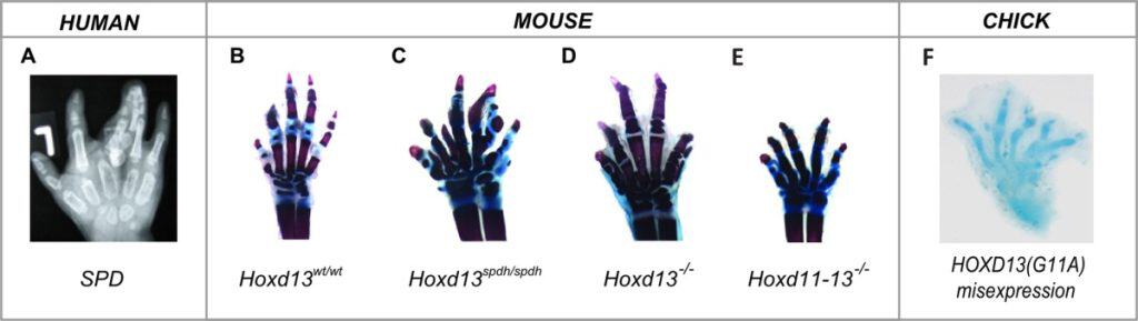
Synpolydactyly primarily affects:
- Distal limb skeletal elements
- Digit formation
- External genital development
Patients with SPD may exhibit:
- Extra digits
- Enlarged skeletal structures
- Limb fusion abnormalities
- Hypospadias
The comparison between HFGS and SPD demonstrates that HOXA13 and HOXD13 may regulate both shared and independent developmental pathways.
Polyalanine Expansions and Protein Dysfunction
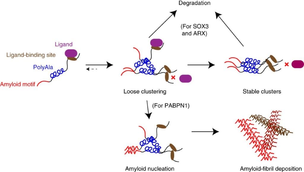
Many HOXD13 mutations involve abnormal expansions of polyalanine tracts within the protein structure. These expansions can alter:
- Protein folding
- Protein-protein interactions
- DNA-binding specificity
- Transcriptional regulation
The severity of developmental abnormalities often correlates directly with the size of the polyalanine expansion.
Researchers propose several mechanisms for mutant HOXD13 activity:
- Disruption of the normal HOXD13 transcriptional network
- General inhibition of genital tubercle development
- Interference with HOXA13-dependent pathways
- Acquisition of abnormal gain-of-function activity
Further molecular investigations are required to fully understand how mutant HOXD13 proteins contribute to congenital genitourinary malformations.
Expression Patterns of HOXA13 and HOXD13
Spatial Distribution During Development
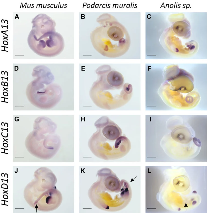
HOX genes typically display spatially organized expression along the anterior-posterior embryonic axis.
Within the genital tubercle, HOXA13 and HOXD13 are among the most highly expressed HOX genes. Both genes show remarkably similar expression patterns, including co-localization within:
- Genital tubercle mesenchyme
- Central epithelial signaling regions
This overlapping expression strongly suggests functional cooperation and redundancy between HOXA13 and HOXD13 during external genital development.
Functional Redundancy and Genetic Interactions
Studies using compound mutant mouse models demonstrate that combined loss of Hoxa13 and Hoxd13 dramatically worsens genitourinary abnormalities.
Observed defects include:
- Severe genital malformations
- Urogenital agenesis
- Urethral closure failure
- Tissue fusion abnormalities
Complete loss of both genes can result in total absence of genital tubercle formation, demonstrating their essential developmental functions.
Human patients carrying combined HOXA13 and HOXD13 mutations also exhibit:
- Reduced labia majora formation
- Recurrent urinary tract infections
- Severe genital abnormalities
These findings confirm that both genes cooperate in regulating normal genitourinary organogenesis.
Cellular Effects of HOXA13 Loss
Regulation of Programmed Cell Death
One of the most important developmental functions of HOXA13 involves regulation of programmed cell death (apoptosis).
During normal genital tubercle development, programmed cell death facilitates:
- Tissue remodeling
- Urethral fusion
- Morphological shaping
- Elimination of transient structures
In Hoxa13 mutant mice, apoptosis is significantly reduced, especially in medial genital mesenchymal regions responsible for tissue fusion.
This defect is associated with loss of BMP7 expression, suggesting that HOXA13 regulates apoptosis partly through BMP signaling pathways.
Regulation of Cell Proliferation
HOXA13 also controls cellular proliferation within the developing genital tubercle.
Loss of Hoxa13 function causes major reductions in cell proliferation due to decreased expression of FGF8 within the urethral plate epithelium.
Experimental supplementation with FGF8 restores proliferative activity in mutant tissues, indicating a strong functional relationship between HOXA13 and FGF signaling during genital morphogenesis.
This interaction represents an important developmental mechanism controlling external genital growth and structural patterning.
Transcriptional Hierarchies in Genitourinary Development
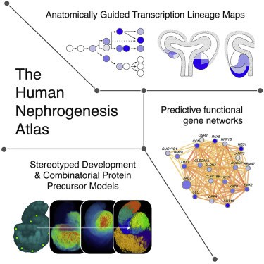
Although significant progress has been made in understanding developmental signaling pathways, relatively little is known about the transcriptional hierarchies controlling genitourinary organ formation.
Future research should focus on:
- Identification of HOXA13 and HOXD13 target genes
- DNA-binding sequence analysis
- Chromatin regulatory mechanisms
- Epigenetic modifications
- Protein-protein interaction networks
- Gene regulatory cascades
Understanding these transcriptional mechanisms may provide critical insights into congenital genitourinary disorders such as:
- Hypospadias
- Vesicoureteral reflux
- Urethral malformations
- Bladder developmental defects
Epigenetic Influences on Genitourinary Malformations
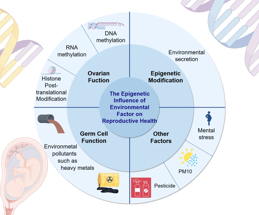
The increasing incidence and diversity of congenital genitourinary defects suggest that environmental and epigenetic factors may also influence embryonic development.
Potential epigenetic mechanisms include:
- DNA methylation abnormalities
- Histone modification changes
- Chromatin remodeling disruptions
- Altered transcription factor accessibility
These epigenetic alterations may interfere with normal HOX protein function and developmental signaling pathways.
Understanding how environmental and epigenetic factors affect genitourinary development could eventually help prevent congenital malformations and improve early therapeutic strategies.
Conclusion
HOXA13 and HOXD13 are critical transcription factors involved in the regulation of external genital and genitourinary development. These genes coordinate essential developmental processes including tissue patterning, cellular proliferation, apoptosis, epithelial signaling, and morphogenesis within the genital tubercle.
Mutations affecting HOXA13 and HOXD13 cause severe congenital syndromes such as Hand-Foot-Genital Syndrome, Guttmacher Syndrome, and Synpolydactyly, all of which can involve major genitourinary abnormalities including hypospadias and urinary tract defects.
Experimental studies demonstrate that these transcription factors function through complex molecular networks involving BMP, FGF, and SHH signaling pathways. Additionally, the overlapping expression and cooperative activity of HOXA13 and HOXD13 reveal important functional redundancy during genital organogenesis.
Continued investigation of HOX gene transcriptional hierarchies, molecular signaling pathways, and epigenetic regulation will be essential for understanding the mechanisms underlying congenital genitourinary malformations and developing future preventive or therapeutic approaches.