
Introduction to Minamata Disease
Minamata Disease is one of the most significant examples of environmental neurotoxicity caused by industrial pollution. The disease was first identified in the 1950s in the coastal region of Minamata after large numbers of residents developed severe neurological disorders following the consumption of seafood contaminated with methylmercury.
The contamination originated from industrial wastewater released by a nearby chemical factory involved in the production of acetaldehyde and vinyl compounds. Methylmercury accumulated in fish and shellfish within Minamata Bay and subsequently entered the human food chain through dietary exposure.
A second outbreak later occurred in Niigata during the 1960s. Unlike the first epidemic, many patients in Niigata presented with mild or subclinical symptoms, making diagnosis more challenging. Over time, thousands of confirmed cases were officially recorded in multiple Japanese prefectures, with a high mortality rate among affected individuals.
Environmental investigations demonstrated extremely elevated mercury concentrations in marine organisms from Minamata Bay. In response, strict mercury regulation policies and large-scale environmental remediation programs, including dredging contaminated sediments, were implemented to reduce mercury exposure and prevent further poisoning.

Methylmercury Toxicity and Disease Mechanism
Mercury as a Neurotoxic Compound
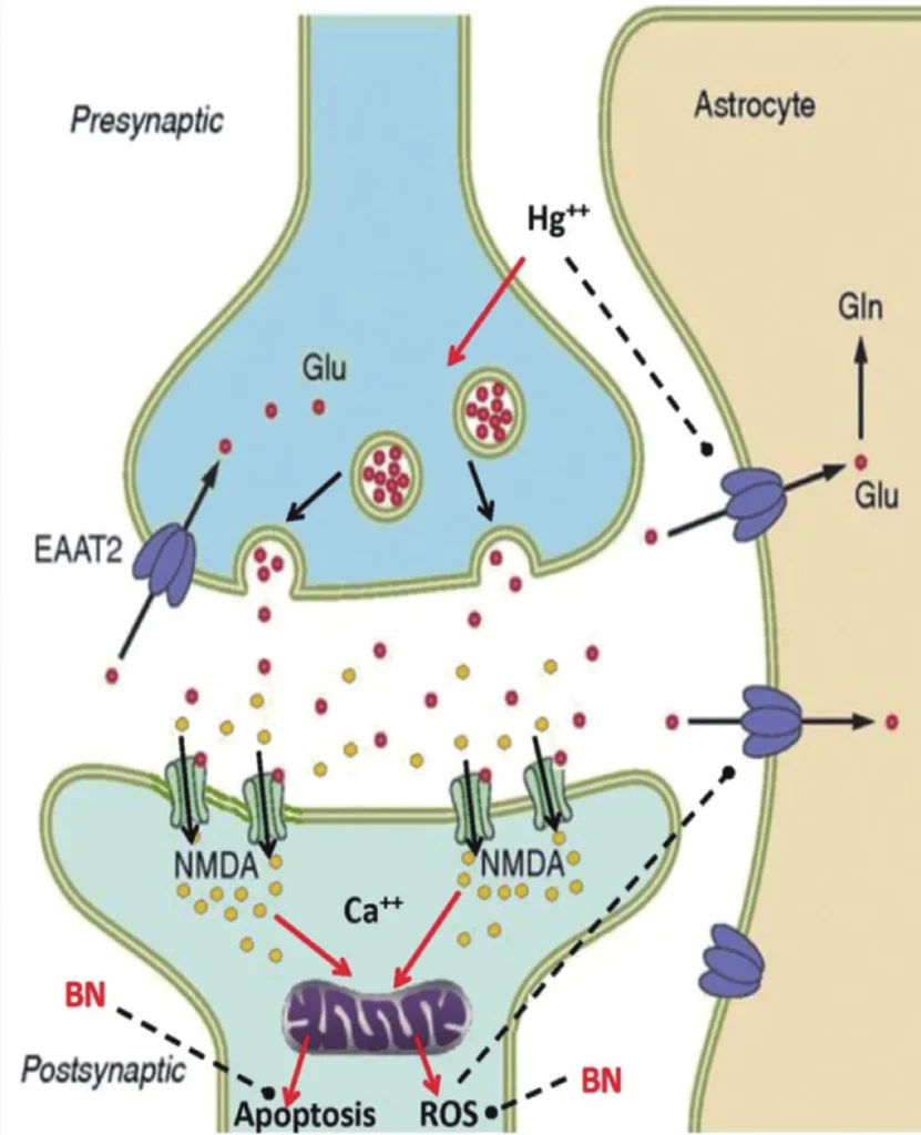
Methylmercury is an organic mercury compound with strong neurotoxic properties. Due to its high lipid solubility, methylmercury easily crosses biological membranes, including the blood-brain barrier and placental barrier. This property allows the compound to accumulate in the central nervous system and fetal tissues.
Inside neural tissue, methylmercury disrupts multiple cellular processes, including:
- Protein synthesis
- Microtubule organization
- Mitochondrial function
- Oxidative stress regulation
- Synaptic transmission
- Neuronal membrane stability
Neuronal injury is particularly severe in regions containing highly metabolically active neurons. Small neurons with limited ribosomal reserves appear especially vulnerable to methylmercury toxicity.
The pathological lesions observed in Minamata disease are highly selective and mainly affect specific cortical and cerebellar regions rather than producing diffuse brain injury.
Clinical Features of Minamata Disease
Neurological Manifestations
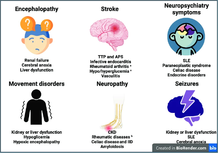
The classical neurological presentation of Minamata disease is commonly referred to as the Hunter–Russell syndrome, characterized by:
- Ataxia
- Dysarthria
- Constriction of the visual field
Additional neurological abnormalities include:
- Sensory disturbances in the extremities
- Disequilibrium
- Tremors
- Muscle weakness
- Hearing impairment
- Abnormal eye movements
- Gait abnormalities
- Taste and smell disturbances
- Psychiatric and cognitive dysfunction
In severe cases, patients may develop paralysis, coma, and death.
Children and fetuses exposed during development often present with more severe neurological damage compared with adults because the immature nervous system is highly sensitive to methylmercury toxicity.
Neuropathology of Minamata Disease
Cerebral Cortex Lesions
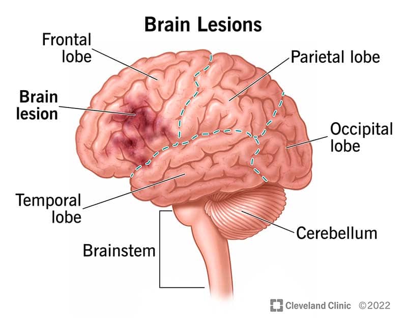
The most characteristic pathological findings of Minamata disease occur within the cerebral cortex. Autopsy studies demonstrated extensive neuronal degeneration, cortical atrophy, and spongiform degeneration in selective cortical regions.
The most severely affected areas include:
- Calcarine cortex
- Precentral gyrus
- Postcentral gyrus
- Temporal transverse cortex
Histopathological alterations include:
- Neuronal swelling
- Acute neuronal shrinkage
- Ischemic neuronal changes
- Neuronophagia
- Perivascular edema
- Demyelination
- Astrocyte proliferation
- Diffuse neuronal loss
Advanced disease may progress to severe cortical atrophy and spongy degeneration of brain tissue.
A grading system was later developed to classify lesion severity, ranging from mild neuronal loss to extensive macroscopic spongiform destruction.
Cerebellar Damage
The cerebellum is another major target of methylmercury toxicity. One hallmark feature is the selective loss of granule cells while Purkinje cells remain relatively preserved during early stages.
Characteristic cerebellar changes include:
- Granule cell depletion
- Purkinje cell disorganization
- Bergmann glial proliferation
- Apical scar formation
- Cerebellar spongiosis
These abnormalities contribute directly to ataxia and coordination impairment observed clinically.
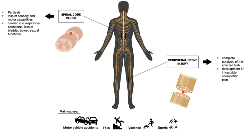
Spinal Cord and Peripheral Nerve Injury
Although primary spinal cord lesions are uncommon, secondary degeneration frequently develops due to cortical neuronal damage.
Peripheral nerve pathology includes:
- Demyelination
- Axonal degeneration
- Abnormal nerve regeneration
- Schwann cell proliferation
- Degeneration of sensory fibers
Sural nerve biopsies from chronic patients revealed incomplete regeneration with irregular branching and small regenerating fibers.
Sensory nerves are generally more vulnerable than motor nerves in methylmercury poisoning.
Fetal Minamata Disease
Prenatal Methylmercury Exposure
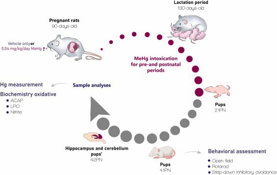
Prenatal exposure to methylmercury results in congenital or fetal Minamata disease, one of the most devastating consequences of environmental mercury contamination.
Methylmercury readily crosses the placenta and accumulates in fetal tissues. In fetal cases, pathology differs from adult disease because the developing brain undergoes disrupted maturation rather than simple neuronal destruction.
Major pathological findings include:
- Neuronal hypoplasia
- Developmental arrest
- Impaired neuronal migration
- Cerebral hypoplasia
- Abnormal cerebellar development
- Defective myelination
Affected infants often exhibit symptoms resembling cerebral palsy, including severe intellectual disability, motor dysfunction, and developmental delay.
Unlike adults, fetal lesions are more diffuse and less restricted to selective cortical regions.
Histochemistry and Detection of Mercury
Histological Identification of Mercury Deposits
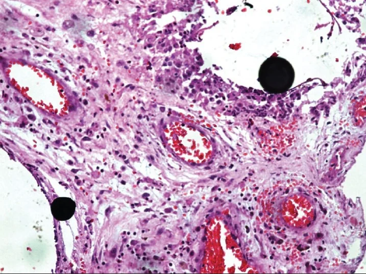
Histochemical techniques have been essential for identifying mercury accumulation in tissues affected by Minamata disease.
Mercury deposits are mainly detected in:
- Kidney proximal tubules
- Hepatic parenchymal cells
- Kupffer cells
- Neurons
- Glial cells
- Perivascular brain regions
Advanced photoemulsion techniques significantly improved sensitivity for mercury detection in pathological samples.
Electron microscopy studies demonstrated that mercury accumulates primarily within lysosomes and is frequently associated with selenium deposition.
Different patterns of mercury accumulation in the brain have been described:
- Neuronal type
- Phagocytic type
- Mixed type
- Perivascular type
These patterns provide insight into mercury transport, metabolism, and cellular detoxification mechanisms.
Organic vs Inorganic Mercury Poisoning
Differential Pathology
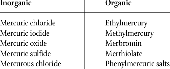
Organic and inorganic mercury poisoning produce distinct pathological and clinical profiles.
Organic Mercury Poisoning
Associated mainly with methylmercury exposure:
- Severe central nervous system injury
- Selective cortical lesions
- Cerebellar degeneration
- Peripheral neuropathy
- Visual impairment
- Sensory deficits
Inorganic Mercury Poisoning
Typically associated with mercury vapor or mercury salts:
- Gingivitis
- Tremor
- Behavioral abnormalities
- Renal toxicity
- Less obvious CNS lesions under routine microscopy
Electron microscopy demonstrates important ultrastructural differences between the two toxicities, particularly involving cytoplasmic inclusions and organelle preservation.
Pathogenesis of Minamata Disease
Selective Neuronal Vulnerability
The precise pathogenesis of Minamata disease remains incompletely understood. However, several mechanisms are believed to contribute to selective neuronal injury:
- Inhibition of protein synthesis
- Oxidative stress induction
- Cytoskeletal disruption
- Impaired axonal transport
- Disturbance of blood-brain barrier function
- Microvascular ischemia
- Excitotoxicity
Selective neuronal vulnerability explains why lesions repeatedly appear in specific brain regions such as the visual cortex, sensory cortex, motor cortex, and cerebellum.
Secondary degeneration of white matter tracts occurs following primary neuronal destruction.
Environmental and Public Health Importance
Minamata disease remains a landmark example of environmental toxicology and industrial pollution. The outbreak transformed international awareness regarding:
- Heavy metal contamination
- Industrial wastewater regulation
- Food-chain bioaccumulation
- Neurotoxicology
- Environmental pathology
- Public health surveillance
Similar mercury pollution concerns continue to emerge globally, including regions affected by artisanal gold mining and industrial contamination.
Modern research on methylmercury toxicity continues to contribute to environmental medicine, neuropathology, developmental neurobiology, and toxicological risk assessment.
Conclusion
Minamata Disease represents one of the most thoroughly studied examples of chronic organic mercury intoxication. The disease primarily targets the nervous system and produces characteristic pathological lesions in the cerebral cortex, cerebellum, and peripheral nerves.
Methylmercury exposure leads to progressive neurodegeneration through complex molecular and cellular mechanisms involving oxidative injury, impaired protein synthesis, neuronal degeneration, and inflammatory responses.
Histopathological and histochemical investigations have played a central role in understanding mercury distribution, tissue damage, and disease progression. In addition, fetal Minamata disease demonstrated the extreme vulnerability of the developing nervous system to environmental toxins.
The study of Minamata disease continues to provide essential insights into neurotoxicology, environmental pathology, and preventive public health strategies aimed at reducing heavy metal exposure worldwide.